There are a lot of ECG courses out there, and from the landing page most of them look the same. Then you're on a Holter report or in device clinic, there's a wide complex tachycardia (WCT) in front of you, it doesn't look like any example you memorised, and the course you did never really made you work one through. So how do you pick an ECG course that actually makes you better at reading ECGs at work?
(Click to zoom on image)
Answer:
Click to Show Answer
Answer:
Choose the course that teaches you why the ECG looks the way it does. The activation sequence, the vector, the anatomy underneath. Then it should make you use that on a lot of ECGs, with a systematic approach and feedback on your answers.
Pattern recognition is the output, not the method. If a course only teaches you what LBBB “looks like”, you’ll recognise the textbook one and be lost the moment it doesn’t look like the textbook.
Full disclosure: I run ECG in Practice, so read this with that in mind. These are the checks I’d use on any course, ours included.
Why does the choice of ECG course matter?
Because ECG interpretation is harder than it looks, even for doctors.
A 2020 meta-analysis of 78 studies found median ECG interpretation accuracy of 54% before training and 67% after training, across all physician training levels (Cook et al.).
Even cardiologists had a pooled pre-training accuracy of about 75%.
The authors’ conclusion: deficiencies at every training level, even after educational interventions.
The ACC/AHA competence statement treats ECG interpretation as a skill that needs supervised practice to reach and ongoing experience to keep (Kadish et al.).
Read that again. That’s doctors, and the 67% is after training.
So “I’ve done an ECG course” doesn’t mean much on its own. A certificate on the wall isn’t the same as reading a WCT properly at 4pm on a Friday. What matters is how the course teaches.
The shortfall which is rarely talked about
Every one of those studies is in physicians or medical students, in a test setting. None of it is in cardiac physiologists.
Accuracy varied widely across studies, and the authors say that could reflect real differences in training or skill, or just differences in assessment design (Cook et al.).
So treat the numbers as a signal, not a precise measure of our profession. These studies can’t tell us how well the numbers transfer to physiologists.
And none of them tested mechanism-first teaching against pattern-first teaching. What follows is my view from teaching, not a trial result.
First, credit where it’s due
There’s never been more ECG content around, a lot of it is free, and some of it is genuinely excellent. The “spot the diagnosis” posts, the flashcards, the mnemonics. They’re quick, they’re addictive, and they get a lot of ECGs in front of a lot of people. That’s a good thing for everyone learning ECGs.
But a memorised pattern is a poor man’s understanding.
It works right up until the ECG in front of you isn’t the one on the flashcard. Then you’re just matching shapes and hoping.
Pattern recognition is the output, not the method
Every waveform on a 12-lead is the heart’s activation sequence, seen from a fixed set of angles. A lead records a positive deflection when the net activation vector points towards its positive pole, and a negative one when it points away (Kligfield et al.). That’s it. If you understand which bit of the heart activates first, which way the wavefront travels and how fast it gets there, you can work out what the ECG should look like before you’ve seen it.
That’s the test I’d put any course through. Here’s what it looks like in practice.
Why is there a small q wave in V6, and why does it vanish in LBBB?
Normally the septum is activated first from its left side, so the first bit of septal activation travels left to right (Durrer et al.).
That early left-to-right wavefront travels away from the left-sided leads and towards V1. That’s the standard explanation for the small ‘septal’ q often seen in I, aVL, V5 & V6 and the small initial r in V1.
In LBBB the left bundle doesn’t conduct. The septum is activated from the right side instead, right to left, and the LV is then activated late and slowly, muscle cell to muscle cell (Strauss et al.).
So the septal q in I, V5 & V6 disappears (absent q waves there are part of the standard LBBB criteria), V1 shows a QS or only a tiny initial r, and the QRS gets broad because the activation is crawling across crappy myocytes not travelling down the slick large cell His-Purkinjie system.
You can memorise “no septal q in LBBB”. Or you can know why. What sort of ECG interpreter do you want to be?
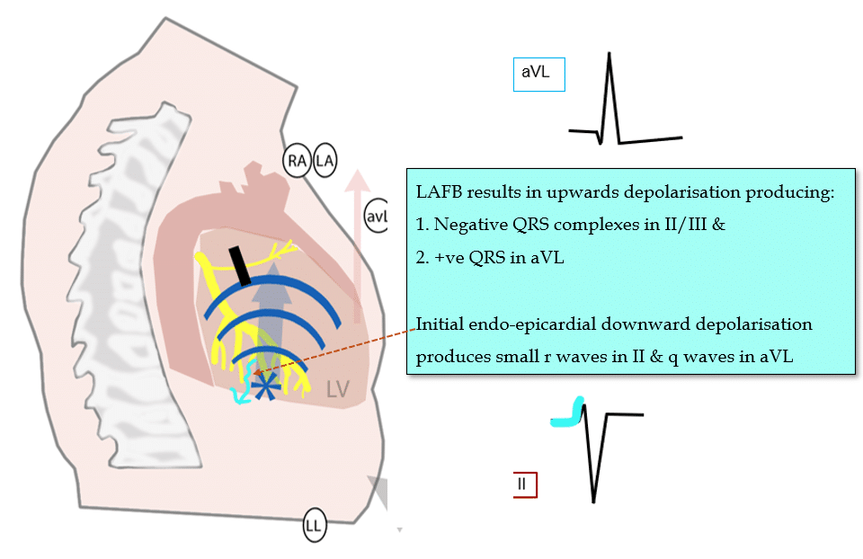
Why does left anterior fascicular block swing the axis up and left?
Block the anterior fascicle and the LV gets activated first through the posterior fascicle, low and inferior (Elizari et al.).
The wavefront then spreads upwards and leftwards to reach the territory the anterior fascicle should have covered.
So the net frontal vector points up and left (Elizari et al.). That’s why the standard criteria include a left axis qR in aVL, and a QRS under 120 ms, because the rest of the conduction system is still doing its job (Surawicz et al.).
Understand where the activation starts makes sense rather than needing to be memorised.
Left anterior fascicular block: the LV activates from below upwards, so II and III go negative and aVL positive.
Why does the start of the QRS help in WCT?
In SVT with aberrancy, the impulse still comes down the His-Purkinje system, so initial ventricular activation is fast. The QRS gets wide later, in the blocked bundle’s territory.
In most VT the impulse starts in the myocardium and spreads cell to cell, so the start of the QRS is slow.
That’s the reasoning behind the lead aVR algorithm’s look at the initial r or q (wider than 40 ms favours VT) and the initial vs terminal ventricular activation velocity (Vereckei et al.).
If you know why the criteria exist, you’re not reciting an algorithm. You’re testing a mechanism. And you’ll know what to do when a criterion is borderline, which it often is.
Why does an outflow tract VT have an inferior axis?
The outflow tracts sit high in the heart. A VT starting there activates the ventricles from the top down, so the frontal vector usually points inferiorly and II, III & aVF are positive (Cronin et al.).
The RV outflow tract is the most anterior part of the heart, sitting right under V1 and V2. A VT starting there sends its wavefront backwards and downwards, away from V1. So V1 is mainly negative (LBBB-like), and the precordial leads turn positive late, usually at V4 or beyond (Cronin et al.).
The aortic cusps and LV outflow tract sit further back, behind the RVOT. From there, more of the wavefront travels forwards, towards the precordial leads. V1 can still look LBBB-like, but the R wave grows earlier and the transition tends to come at or before V3 (Cronin et al.).
VT localisation from the QRS is anatomy plus activation sequence. Nothing more mysterious than that.
The sad reality is that plenty of people can name these patterns and can’t tell you why a single one of them looks the way it does. Is the bar really so low for Cardiac Physiologists?
How much practice & feedback do you get?
You don’t learn ECGs by watching someone else read them. It feels like learning. It isn’t.
Look for lots of real ECGs to interpret yourself, with worked answers that explain the mechanism, not just the diagnosis.
Look for assessment. A review of ECG teaching methods found self-directed learning was associated with poorer competence, and that summative assessment improved performance (Fent et al.).
Check whether the cases are graded from easy to hard. Being thrown straight into complex tracings is discouraging. Staying on easy ones forever is pointless.
Being told “correct” without the reasoning isn’t knowing something. You need to know why you were right, or you’ll be wrong the next time it looks slightly different.
Does it go beyond the basics?
Most courses cover sinus rhythm, blocks & a STEMI. Fewer go deep on the ECGs that actually cause trouble, and that’s exactly where a list of patterns runs out.
Questions to ask:
Does it teach WCT properly, including VT vs aberrancy, and why each criterion works?
Does it build VT localisation from the anatomy? Where the VT starts sets the activation sequence, and the activation sequence sets the axis, the bundle branch block shape and the precordial transition.
Does it handle the “looks like X, is actually Y” ECGs, e.g. isorhythmic AV dissociation vs complete heart block? In isorhythmic AV dissociation the two rates are almost the same, so the P waves drift in and out of the QRS, and a P wave that lands far enough ahead of the QRS can still conduct.
Does it connect conduction disease to what happens next for the patient? That means working out where the block is (in the AV node or below it), why that changes how urgent it is, and when an EP study or pacemaker comes into it (Kusumoto et al.).
If the course stops at the basics, it may suit a student. It probably won’t stretch a working physiologist. And the basics aren’t what keeps you up at night after a Holter report.
Is it built for cardiac physiologists?
A course written for medical students or nurses can still be good. But physiologists read ECGs in differently. Medical students need the high yield pneumonic, recognise and move on. For most, ECG is all stress and little love. Trust me, I’m in Med School now. That’s no dig on them either. The sheer volume, breadth and pace of the content their learning is immense. They cant get caught up in P wave localisation or inverted sinus p waves. But Cardiac Physiologists can. Theres no excuse for being the lowest standard possible for your job.
Do the examples look like what you see at work?
Does it explain the mechanism, not just the criteria?
Does it link the surface ECG to what you’d see on intracardiac EGMs, if you work in EP or plan to? It’s the same activation sequence, seen from a different place.
If every example looks like it came from an emergency department, it probably wasn’t written with you in mind.
Online or face-to-face?
Both can work. The 2022 meta-analysis found a small advantage for computer-assisted over face-to-face instruction, and found group study did better than studying alone (Oh et al.).
So an online course is fine, but ideally with some way to discuss cases with others or ask questions, rather than working completely alone.
What about access & support?
How long do you keep access? ECG skills fade without use, and you’ll want to come back.
Can you ask a question and get an answer from someone who knows?
Is there a certificate or progress record if your department needs evidence of training?
Any structured course beats chance exposure
Of course I think ECG in Practice is a good option. But the real point is, you should commit to structured learning, even if it’s not ECG in Practice. Turning up and hoping you’ll pick ECGs up along the way isn’t a system.
If your department’s plan is essentially “expose & hope for the best”… that’s not a system. That’s chance.
And if you’re heading for the EP lab after this, it’s the same idea, just deeper. More than 130 students across 15 countries train with EP in Practice, which is over 100 hours across 8 programs.
My two cents:
Pick the course that teaches you why the ECG looks the way it does, not the one that hands you the most patterns to memorise.
Then make sure it drills a systematic approach. That has the strongest evidence behind it.
Check the depth. WCT, VT localisation & conduction disease separate a physiologist-level course from an intro course, and every one of them is a mechanism question.
The sad reality is that plenty of people do an ECG course, tick the box, memorise a few more shapes, and read ECGs exactly the way they did before. Minimum standard. Lots of experience doing a crap job of reading ECGs really doesn’t count for much. Don’t pick the course that lets you do that.
Thanks for tuning in :)
Cheers Mitch & CPiP Team
A quick checklist
[ ] It explains why each waveform looks the way it does: the activation sequence, the vector, the anatomy
[ ] A systematic approach you’re made to use on every ECG
[ ] Lots of ECGs to interpret yourself, with worked answers that explain the mechanism
[ ] Assessment, not just reading
[ ] Depth beyond the basics, taught as mechanism: WCT and VT vs aberrancy, VT localisation, conduction disease
[ ] Examples from the settings physiologists work in
[ ] A clinically active author & referenced content
[ ] Someone to ask, and access long enough to revisit
If you want to see how ECG in Practice is set up, the ECG in Practice page lists what’s covered.
FAQ
What should an ECG course for cardiac physiologists include?
Teaching that explains why each waveform looks the way it does (the activation sequence, the vector and the anatomy), a systematic interpretation approach, many real ECGs to interpret with worked answers and assessment, and depth beyond the basics, including wide complex tachycardia (WCT), VT vs aberrancy, VT localisation and conduction disease, using examples from settings physiologists work in.
Is pattern recognition enough to read ECGs?
Not on its own. Patterns are the result of the heart’s activation sequence. If you understand the mechanism, you can work out an unfamiliar ECG. If you’ve only memorised the pattern, you can only match what you’ve already seen.
Are online ECG courses as effective as face-to-face teaching?
The evidence suggests they can be. A 2022 meta-analysis found a small advantage for computer-assisted instruction over face-to-face teaching, and better results with group study than studying alone.
What is the best way to improve ECG interpretation?
Use a systematic approach on every ECG, ask why each finding looks the way it does, interpret lots of ECGs yourself, and get feedback or assessment on your answers. Systematic approaches showed a large effect in a 2022 meta-analysis of ECG training.
Is one ECG course enough?
Usually not. Accuracy improves with training but stays imperfect at every level, so ongoing practice matters as much as the course itself.
References
Cook DA, Oh SY, Pusic MV. Accuracy of physicians’ electrocardiogram interpretations: a systematic review and meta-analysis. JAMA Intern Med. 2020;180(11):1461-1471. doi:10.1001/jamainternmed.2020.3989. PMID 32986084.
Oh SY, Cook DA, Van Gerven PWM, Nicholson J, Fairbrother H, Smeenk FWJM, Pusic MV. Physician training for electrocardiogram interpretation: a systematic review and meta-analysis. Acad Med. 2022;97(4):593-602. doi:10.1097/ACM.0000000000004607. PMID 35086115.
Fent G, Gosai J, Purva M. Teaching the interpretation of electrocardiograms: which method is best? J Electrocardiol. 2015;48(2):190-193. doi:10.1016/j.jelectrocard.2014.12.014. PMID 25573481.
Kadish AH, Buxton AE, Kennedy HL, et al. ACC/AHA clinical competence statement on electrocardiography and ambulatory electrocardiography. Circulation. 2001;104(25):3169-3178. doi:10.1161/circ.104.25.3169.
Kligfield P, Gettes LS, Bailey JJ, et al. Recommendations for the standardization and interpretation of the electrocardiogram: part I: the electrocardiogram and its technology. Circulation. 2007;115(10):1306-1324. doi:10.1161/CIRCULATIONAHA.106.180200.
Durrer D, van Dam RT, Freud GE, Janse MJ, Meijler FL, Arzbaecher RC. Total excitation of the isolated human heart. Circulation. 1970;41(6):899-912. doi:10.1161/01.cir.41.6.899.
Strauss DG, Selvester RH, Wagner GS. Defining left bundle branch block in the era of cardiac resynchronization therapy. Am J Cardiol. 2011;107(6):927-934. doi:10.1016/j.amjcard.2010.11.010.
Surawicz B, Childers R, Deal BJ, Gettes LS. AHA/ACCF/HRS recommendations for the standardization and interpretation of the electrocardiogram: part III: intraventricular conduction disturbances. Circulation. 2009;119(10):e235-e240. doi:10.1161/CIRCULATIONAHA.108.191095.
Vereckei A, Duray G, Szénási G, Altemose GT, Miller JM. New algorithm using only lead aVR for differential diagnosis of wide QRS complex tachycardia. Heart Rhythm. 2008;5(1):89-98. doi:10.1016/j.hrthm.2007.09.020.
Brugada P, Brugada J, Mont L, Smeets J, Andries EW. A new approach to the differential diagnosis of a regular tachycardia with a wide QRS complex. Circulation. 1991;83(5):1649-1659. doi:10.1161/01.cir.83.5.1649.
Cronin EM, Bogun FM, Maury P, et al. 2019 HRS/EHRA/APHRS/LAHRS expert consensus statement on catheter ablation of ventricular arrhythmias. Heart Rhythm. 2020;17(1):e2-e154. doi:10.1016/j.hrthm.2019.03.002.
Kusumoto FM, Schoenfeld MH, Barrett C, et al. 2018 ACC/AHA/HRS guideline on the evaluation and management of patients with bradycardia and cardiac conduction delay. Circulation. 2019;140(8):e382-e482. doi:10.1161/CIR.0000000000000628.