Localising VT using the QRS morphology

Question:

Assuming this is VT, where is this VT arising from?

(Click to zoom on image)

Answer:

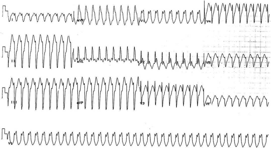
Answer: Inferoapical Left Ventricle (not far from Posteromedial Papillary Muscle)


Features indicating an LV origin

  • Earlier precordial transitions (meaning, the earliest evidence of a dominantly positive precordial lead) indicate an LV origin with increasing likelihood the earlier the precordial transition.
    • Regarding precordial transition & chamber of origin I would usually say:
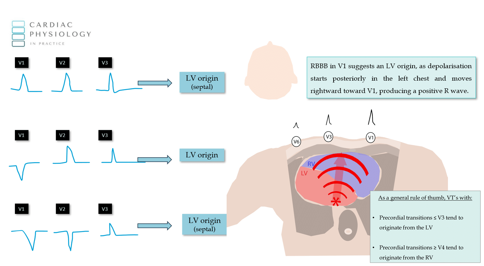
      • “If V1 is positive, you can bet your house its originating from the LV”

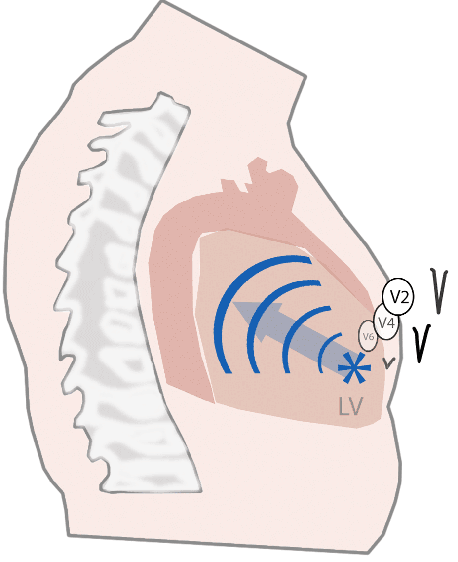
      • “If V2 is the first dominantly positive QRS, then you can bet your car on an LV origin”

      • “If V3 is the first dominantly positive QRS, then…don’t bet anything you care about, but, gun to your head with no other information, bet on the LV”


  • In this ECG, precordial transition is as early as it can get (V1 is positive), we can bet our house that this VT is exiting within the left ventricle. (Please do not take financial advice from me!).


Features indicating an Apical origin

  • Since the majority of the precordial leads are negative, the exit site for this VT is likely located closer to the apex than the base of the heart. This is because an apical originating VT will depolarise the heart AWAY from the chest wall (and hence, away from the precordial leads), producing a negative deflection in the precordial leads V3-V6.


  • Determining basal vs apical origin is important, because it provides clues on the mechanism of VT. Most (but not all) idiopathic VT’s originate from the base of the RV or LV in the RVOT or LVOT. An apical VT makes an idiopathic cause less likely, and may suggest a scar related mechanism (although not always – consult your EP!).


Features indcating an inferior wall origin

  • Lead II/III/aVF are all negative, indicating an inferior wall origin.


  • Depolarisation initiating from the inferior wall of the LV, depolarises the heart in an upward direction.


  • This vector of depolarisation “opposes” the vector of II which goes from the RA –> LL, hence lead II is negative in this example.


Endocardial or Epicardial origin?
    • Another interesting point, is that we can surmise that this VT originates from the endocardium, and not the epicardium. This is important, because it may mean it can be treated with ablation & can give clues about the aetiology of the VT since some diseases/cardiomyopathies have propoensities for generating epicardial vs endocardial VT’s.


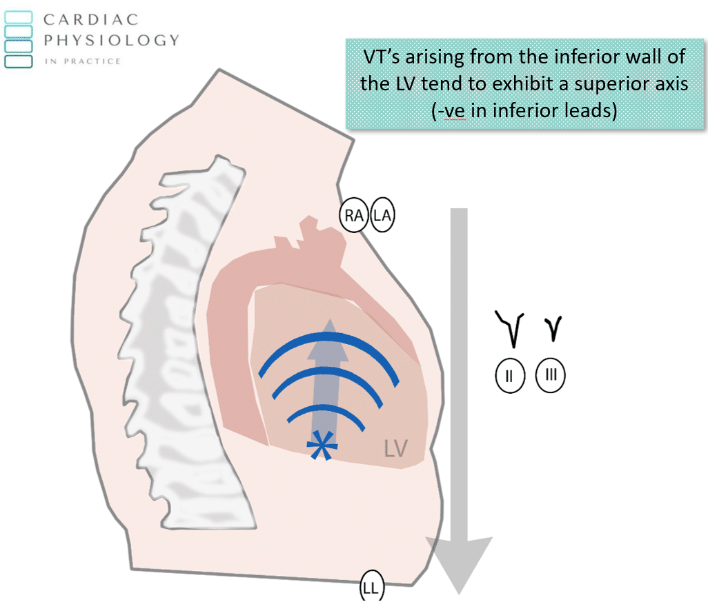
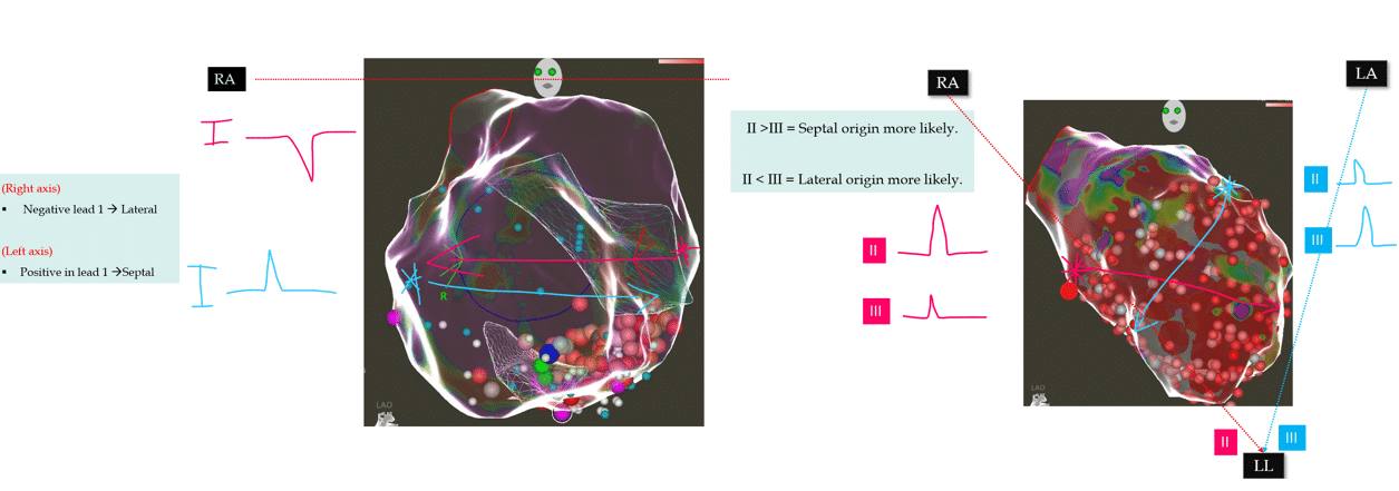
    • The small “r” wave in the inerior leads can be observed due to initial endocardial to epicardial depolarisation. This transmural conduction moves downwards, producing a small r wave in the inferior leads in VT’s arising from the inferior wall endocardium.


      • VT’s arising from the epicardial cardiac crux region for example, often lack this initial “r” wave in the inferior leads.


Features indicating Septal Vs Lateral Origin. 

  • Lead 1 is mostly negative, indicating a lateral wall origin, however, since the apex is a laterally located structure, this may also account for the predominantly negative lead 1.


  • The small “r” wave in lead 1 does indicate that at least some depolarisation is moving from right to left (WITH the trajectory of the Lead 1 vector from RA –> LA). This indicates the possibility of a septal origin, despite a dominantly negative lead 1 morphology.


  • If the lead II is (on average) more positive than lead III (or less negative), then this also indicates a more septal origin, since the lead II vector has a leftward swing to it (RA –> LL), compared to the lead III vector which moves mostly down and slightly left (LA –> LL).


My Two Cents

  • Localising the approximate origin of VT can be readily performed once you understand cardiac anatomy & the ECG vectors.
  • Any depolarisation that moves with the ECG vectors, produces a positive deflection and vice versa.
  • In this case, a positive deflection in V1 (a right sided anterior chest lead), indicates an origin in the left posterior chest, hence indicating an LV origin.
  • A “superior axis” (negative QRS in II/III/aVF) indicates an inferior wall origin, with the addition of the small initial “r” wave indicating an endocardial origin.
  • Negative precordial leads indicate depolarisation starting near the precordial leads (anterior chest, where the APEX is) and moving away.
  • Lead 1 is dominantly negative, usually indicating a lateral wall origin, however, the small r wave initially indicates a possible septal origin.


Thanks for tuning in :)
Cheers
Mitch & CPP Team


    • VT localisation is discussed in far more depth in: ECG in Practice & EP in Practice.
    • Additional advanced features of VT localisation are discussed in EP in Practice.

Leave a Reply

Subscribe to stay informed

Get the latest updates and event details, and be notified when new courses launch.