VT ablation using Decrement Evoked Potential (DeEP) mapping

Question:

These isochronal maps represent the same ventricle, both mapped during RV pacing. How can they be so different?

(Click to zoom on image)

Answer:

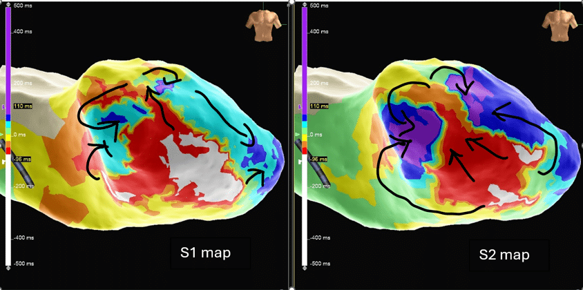
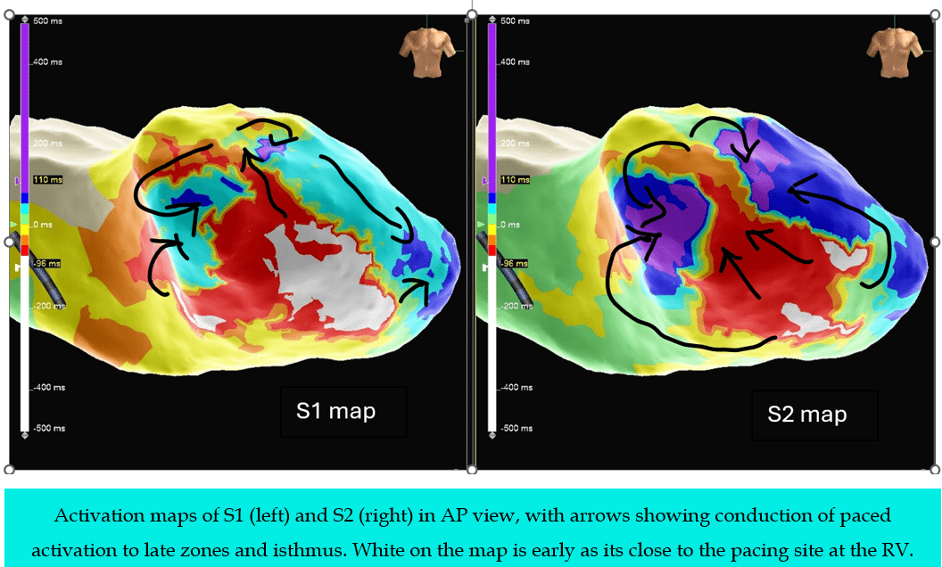
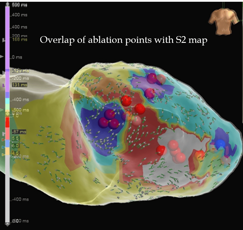
Answer: One was mapped during S1 (Drive train), the other was mapped during S2 (Extra Stimulus)


VT Ablation & DeEP Mapping concepts

  • With advances in technology, eg multi-electrode mapping and automated mapping, there is a far better understanding of VT circuits in recent years. VT ablation progressed from scar homogenization to isochronal late activation mapping (iLAM) and recently Decrement Evoked Potential (DeEP) mapping.


  • DeEP mapping uses S1 S2 continuous pacing throughout the mapping phase. The shorter S2 CL from S1 further emphasizes the areas of not only slow conduction but areas which decrement.


  • The requirements for re-entry are unidirectional block and slow, decrementing areas, which can be critical in initiating and maintaining circuits.


  • DeEP mapping is a method of “unmasking” these regions of functional conduction slowing which may critically participate in VT circuits by pacing a rapid CL on S2 & noting any late potentials observed.


Methodology of DeEP Mapping

  • DeEP mapping would be done by firstly, determining the Ventricular Threshold and Ventricular ERP. With these results, RV pacing was done at these settings, S1S2 CL (cycle length) at S1 600ms (or higher than patient’s intrinsic rhythm), with a shorter drive of 4-6 beats instead of the usual 8 beats for EP study to facilitate faster mapping. For S2, CL would be set at 30ms > VERP. This pacing sequence is repeated until map is complete.


  • Areas of isochronal crowding are already present on the S1 map, the side by side comparison shown below demonstrates the late areas and areas of isochronal crowding is further emphasized and better defined on the S2 map.


  •  In this particular case, VT induction then produced a haemodynamically stable VT allowing us to map the VT circuit.


  • From the video, we can see that the isthmus of VT & the isthmus of late potentials identified on the S2 map coincide. Moreover, positioning the HD grid at this isthmus site during VT, showed mid diastolic potentials.


Ablation Termination

  • HD grid positioned at site of S2 decremental late potentials yielded Mid Diastolic signals during VT. Although this does not confirm participation of this tissue during VT, it is highly suggestive of it, especially as the VT propogation map suggests the VT isthmus was in this region.


  • Ablation in this region terminated VT, all but proving that this region of decremental S2 Late Potentials during RV pacing (& mid diastolic potentials during VT) were participating as the critical isthmus during VT circuitry.


Summary:

  • DeEP Mapping is performed by mapping signal voltage & timing on S2 where S2 is programmed within 30ms of VERP.


  • DeEP Mapping may uncover regions of diseased issue, by manifesting regions of functional conduction veliocity slowing. These regions of slow conduction may be critically involved in VT circuitry.


  • These regions may conribute to isochronal crowding during RV Pacing rhythm, and represent targets for ablation in modern VT ablation.


Regards

This post, including the images & videos was authored by:


Liang Shufen CEPS, CCDS

Principal Cardiac Physiologist

IBHRE Ambassador

National Heart Centre Singapore


The CPP Team thanks her for her incredibly valuable insights & contributions to Cardiac EP education!

Leave a Reply

Subscribe to stay informed

Get the latest updates and event details, and be notified when new courses launch.