Thinking beyond ST Elevation: Prognosis, bundle branches & septal perforators

Question:

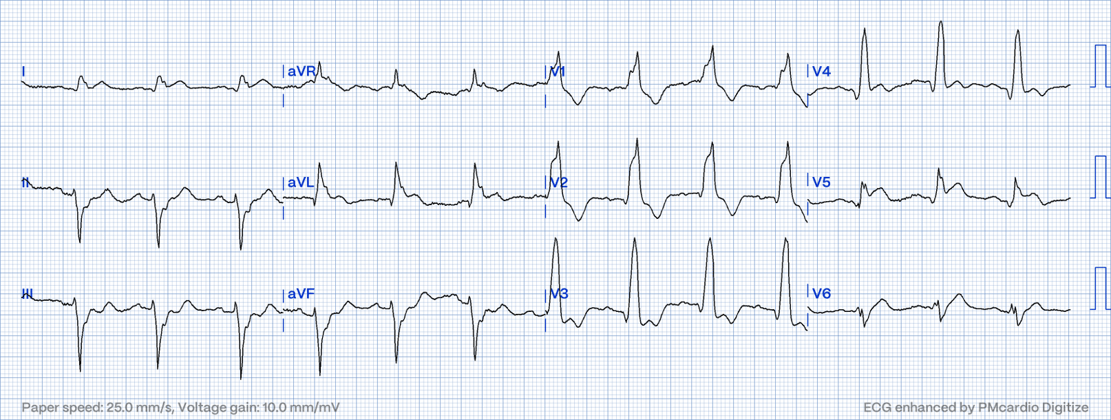
87 year old with resting chest pain with ECG sent in by ambulance. No further information. What is your interpretation of this ECG?

(Click to zoom on image)

Answer:

“This Gentleman had a proximal LAD occlusion resulting in myocardial infarction, treated with emergency PCI in the Cath lab.”

Sinus rhythm with Right Bundle Branch Block (RBBB), Left Anterior Fascicular Block (LAFB), and an occlusive myocardial infarction (OMI) impacting the Left Anterior Descending Artery (LAD), proximal to the 1st Diagonal Branch resulting in anterolateral ischemia.


  • The presence of ST elevation (STE) in leads V4-V6 indicates apical (or lateral) LV ischemia, with STE in leads I & aVL suggesting lateral LV ischemia. Initially, this would raise suspicion of Left Circumflex Occlusion. However, concurrent apical & lateral ischemia most often arises from an occlusion in the proximal LAD, as the LAD supplies the anterior wall & the apical LV. The LAD is also responsible for supplying part of the LV lateral wall (leads I & aVL) via the 1st diagonal branch (D1). ST elevation in aVL typically points to ischemia in the D1 or Obtuse Marginal territories. In combination with anterior or apical ischemia, it is usually associated with a proximal LAD occlusion.


  • Detecting anterior ischemia via the ST segments in V1-V3 is challenging due to the RBBB; however, subtle “concordant” elevation at QRS offset may indicate concurrent anterior ischemia. Certainly v4 demonstrates ST elevation indicating an anterior injury current. Additionally, the possibility of new-onset RBBB and LAFB should be considered, as both components of the conducting system are supplied by the LAD, and a proximal LAD OMI can damage these structures.


  • The emergence of RBBB and LAFB in the context of LAD OMI is associated with a poorer prognosis. Partly, this is because the RBBB is supplied by a very proximal septal perforator branch from the LAD. If the RBB is injured during an anterior MI, it often indicates a very proximal LAD occlusion with a risk of an extensive resulting myocardial injury.


Thanks for tuning in :)
Cheers
Mitch & the CPP Team

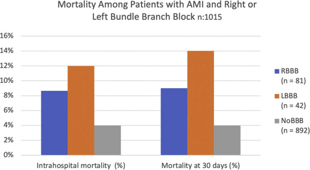
Figueroa-Triana JF, Mora-Pabón G, Quitian-Moreno J, Álvarez-Gaviria M, Idrovo C, Cabrera JS, Peñuela JAR, Caballero Y, Naranjo M. Acute myocardial infarction with right bundle branch block at presentation: Prevalence and mortality. J Electrocardiol. 2021 May-Jun;66:38-42.

One Response

  1. As a person who works in paediatrics, these examples are very helpful to keep sharp on ischaemia/infarction ECG interpretation. 👍 👍 👍

Leave a Reply

Subscribe to stay informed

Get the latest updates and event details, and be notified when new courses launch.