Intramural VT: Highlighting the limitations of “Contact Mapping”

Question:

This patient had a prior inferoseptal–apical myocardial infarction. Endocardial and epicardial activation mapping during VT (CL 330ms) demonstrated nearly simultaneous earliest activation on both surfaces with a notable absence of mid diastolic potentials on both surfaces. Given this information & the maps & EGMs below - what can be said about the location of the VT circuit?

(Click to zoom on image)

Answer:

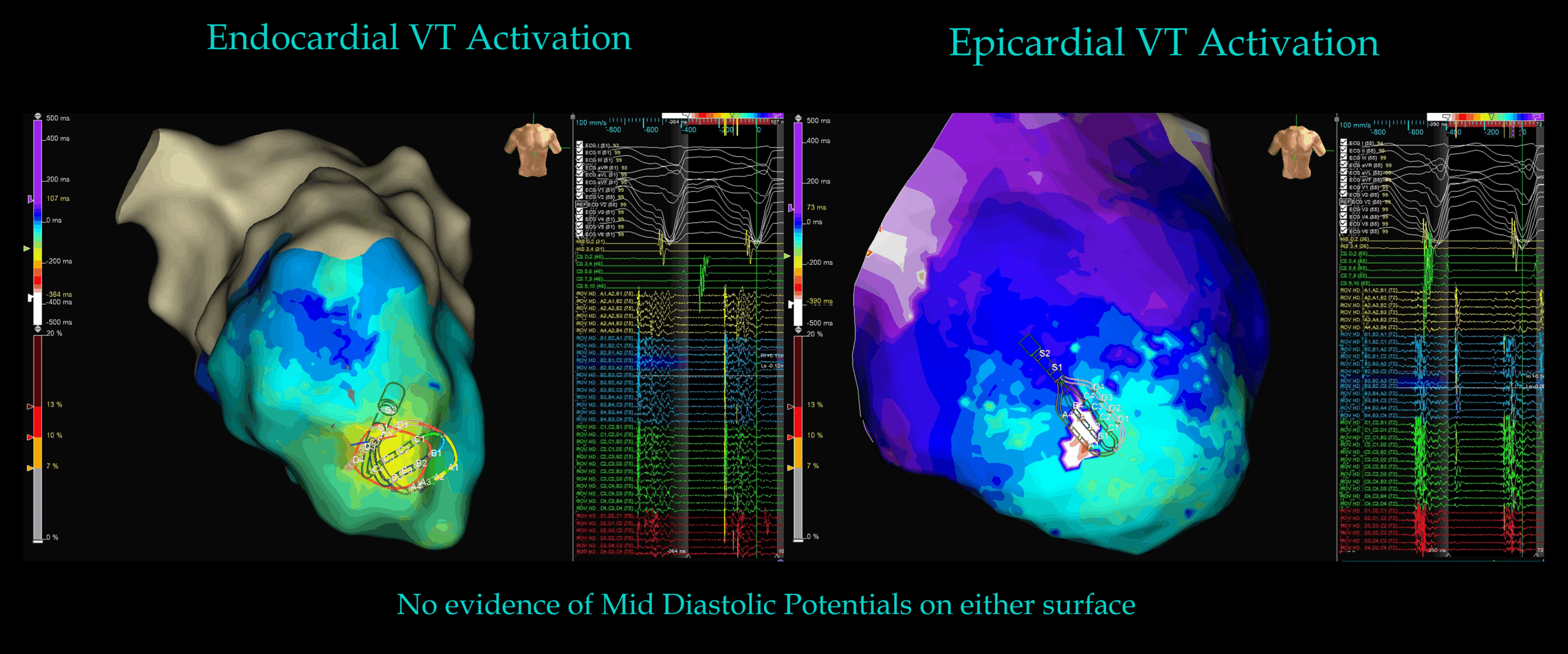
Answer: The mapped sites represented breakout rather than the critical isthmus. Critical isthmus site must be intramural, rather than endo/epicardial.


This case highlights a number of interesting points:

  • Firstly, it highlights that ischaemic VT circuitry can be intramural and we cannot make assumptions that ischaemic VT substrate is mostly endocardial.


  • Secondly, this case highlights that pseudo-focal activation does not exclude reentry. Simultaneous endo/epi activation/breakout strongly suggests the presence of an intramural circuit.
    • Propagation maps confirmed synchronized ENDO–EPI emergence from deeper tissue.


Substrate mapping:

  • Voltage mapping demonstrated dense, heterogeneous scar without a clear endocardial conduction corridor – yet this was unlikely the critical isthmus site for the VT due to the lack of diastolic potentials (figure below).


  • Programmed stimulation (S2 mapping) unmasked LAVAs and late potentials at the inferoseptal apex, with truly marvelous isochronal crowding indicating critically slow conduction —>which is once again predictive of a VT critical isthmus site -yet this was unlikely the critical isthmus site, as demonstrated by the lack of mid diastolic potentials during VT, and the focal breakout pattern revealed during VT activation mapping


  • S2 timing map figure below – indicating late potentials and isochronal crowding.


Ablation targeted both endocardial and epicardial breakout regions, resulting in non-inducibility.


My Two Cents:

  • Ischaemic substrate can be primarily intramural.


  • Late potentials and Iscohronal Crowding are predictive (but not confirmatory) of VT isthmus sites & diastolic corridors – but in this case, they were not part of the VT isthmus site.


  • Traditional “contact mapping” mapping is really only a 2-Dimensional surface map of the endocardium or epicardium – we cannot map intramural circuitry with current technology in the EP lab.


  • Tools such as Unipolar Voltage, ICE, MRI & educated inferences are our best tools for deducing intramural VT circuitry.


  • Ablation at the breakout points was sufficient in abolishing VT in this instance.


Thanks for tuning in :)

Regards

Author: Dr Juan. Ismael Almonte G. (MD) with colleagues Dr Rodolfo San Antonio Dharandas (MD) and Judit Mas, Biomedical Engineer – Abbott / EnSite X Specialist.

Edited by Mitch & CPiP Team with permission.

EP in Practice – Product Overview

REFERENCES

  1. Stevenson WG, Wilber DJ, Natale A, et al. Irrigated radiofrequency catheter ablation guided by electroanatomic mapping for recurrent ventricular tachycardia after myocardial infarction: the multicenter ThermoCool VT trial. Circulation. 2008;118(25):2773-2782.
  2. Jaïs P, Maury P, Khairy P, et al. Elimination of local abnormal ventricular activities: a new end point for substrate modification in patients with scar-related ventricular tachycardia. Circulation. 2012;125(18):2184-2196.
  3. Ciaccio EJ, Akar JG, Ramanathan C, et al. Structure and function of the ventricular tachycardia isthmus in postinfarction reentrant VT. Heart Rhythm. 2022;19(3):439-447.
  4. Al-Khatib SM, Stevenson WG, Ackerman MJ, et al. 2017 AHA/ACC/HRS guideline for management of patients with ventricular arrhythmias. Heart Rhythm. 2018;15(10):e73-e189.
  5. Anter E, Tschabrunn CM, Josephson ME. Ablation of reentry-vulnerable zones in scar-related VT. Circ Arrhythm Electrophysiol. 2020;13(2):e008625.
  6. Ghanbari H, Basu S, Henrikson CA, et al. Noninducibility in post-infarction VT as predictor of outcome. Circ Arrhythm Electrophysiol. 2014;7(3):533-540.
  7. Guandalini G, Liang JJ, Hutchinson MD. Ventricular tachycardia ablation: past, present, future. JACC Clin Electrophysiol. 2019;5(12):1446-1458.
  8. Martin R, Maury P, Shah AJ, et al. Characterization of VT isthmus with high-density mapping. Circ Arrhythm Electrophysiol. 2019;12(11):e007567.
  9. Cronin EM, Bogun FM, Maury P, et al. 2019 HRS/EHRA consensus on catheter ablation of ventricular arrhythmias. Heart Rhythm. 2020;17(1):e2-e154.
  10. Lo R, Chia KKM, Hsia HH, et al. Ventricular tachycardia in ischemic heart disease: mechanisms and ablation evidence. Card Electrophysiol Clin. 2017;9(1):25-46.

Leave a Reply

Subscribe to stay informed

Get the latest updates and event details, and be notified when new courses launch.