PFA was meant to be the safe energy near the AV node. Tissue-selective, and thought to spare the conduction system. So why might it injure the AV node, and why might it not?
(Click to zoom on image)
Answer:
Click to Show Answer
The first slow-pathway pilot put the expectation in writing. Its introduction said the “high tissue selectivity characteristic of PFA could potentially protect nontarget tissue from damage, such as the His bundle.”
Then a 40-patient slow-pathway series reported transient AV block in 17.5%. Most of it was complete heart block. They used a focal PFA catheter in a biphasic/bipolar manner.
So “tissue-selective” needs a closer look. There are reasons the node might fare better, but that isn’t the same as knowing it won’t be injured.
What does “tissue-selective” actually mean?
Electroporation is when an electric field pushes voltage across a cell membrane past a critical level – resulting in irriversible tissue injury.
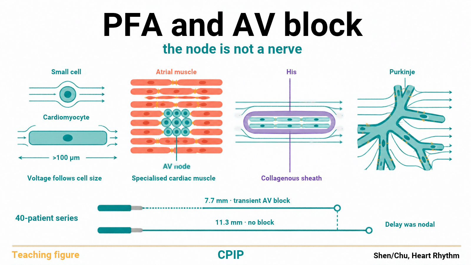
The maximum voltage induced across a cell is proportional to its size. For microsecond pulses, larger cells are more susceptible to ablation than small ones
Working cardiomyocytes are large, elongated cells, often more than 100 µm long.
Nerves by comparison have layered connective-tissue sheaths, often with myelin. Vessels & the oesophagus have layered smooth muscle.
Under identical lab conditions, cardiomyocytes have shown a lower electric field threshold for irriversible electroporation than smooth muscle cells and neurons.
That’s the basis of the selectivity argument. But there are limits:
Not every study agrees. One suspension study found neuronal cell lines and an atrial cardiomyocyte line died at very similar field strengths.
Non comparable catheters & waveform cocktails –> Pulse duration, timing & dosing change it. You can’t take one number from one PFA system and use it for another.
But…the AV node is not a nerve???
No. Neither is the His–Purkinje system.
The selectivity argument usually refers to myocardium vs nerve, vessel & oesophageal tissue. The node is specialised cardiac muscle…so how does PFA interact?
The AV node & slow pathway is made up of smaller sized cells…perhaps making it less susceptible to PFA.
This could raise the threshold for PFA ablation and lend the AV node some protection from PFA.
Duchateau et al.’s 2026 Europace review predicts that cells with a smaller diameter, such as the AV node, may be less susceptible to similar electric field exposure, leading to reversible electroporation rather than irreversible injury. Thats good news for avoiding injuury, however, its possible that surrounding myocardium may still be ablated, potentially isolating the node from the rest of the myocardium. A few more points:
No published study has measured nodal electroporation thresholds.
Coupling, fibre direction & insulating tissue shape the field as well…so the logic becomes real messy real quick.
One proposed mechanism of pFA injury is via inducing calcium overload….that couold make the node MORE susceptible to injury given it’s calcium dependence.
PFA-CONDUCT used monopolar biphasic focal PFA on 3 pigs directly at the AV junction. It caused durable complete AV block in 100% of pigs after 2–3 applications…so clearly the node CAN be ablated with PFA.
However, the same study was unable to permanently injure the AV node when attempting ablation from the NCC (0/5 pigs). So distance, and intervening tissue may distribute voltage & protect the node.
So… on the one hand, PFA is absolutely capable of blocking the node. On the other hand, it has some characteristics which may give us a reason to expect a different ablation threshold vs surrounding structures. We don’t know what a safe dose is yet.
How about the His bundle and the Purkinjie fibres?
Sugrue et al. tested bipolar pulses in a canine study:
The pulses acutely wiped out Purkinje potentials -im not suprised, purkinjie cells are large cells, and therefore theoretically should experience greater voltage per pulse and be more susceptible to PFA mediated injury than surrounding tissue. In saying that…this is not entirely true or clear cut. Some authors have reported durable purkinjie sparing with PFA ablation….so the truth is still kind of unknown.
In the two animals where they tried, they couldn’t alter the His signal, even at 2,250 V. The authors suggested the His bundle’s collagenous sheath changes how the field is distributed and perhaps insulated the His bundle from recieving a high voltage dose.
What do the distance data actually show?
The closer to the His you ablate, the more chance you will injure the His. In a 40-patient series where the authors attempted slow pathway ablation using PFA, the lesions associated with transient AV block were closer to the His.
Lesions with block sat 7.7 mm from the His, compared with 11.3 mm for those without block.
AH delayed first in most, while HV stayed constant. Interestingly, this indicated the AB block mechanism was nodal.
Perhaps the calcium dependence of the node made it more susceptible, or perhaps it was isolated from surrounding myocardium, or hperhaps the His was protected by its fibrous sheath redistributing voltage…sadly we don’t really know. Theres much about PFA we don’t understand.
PR was back to normal within 24 hours.
My two cents:
The selectivity that protects the phrenic nerve & oesophagus was never evidence about the AV node. The node is muscle.
Small nodal cells and the His bundle’s sheath give us reasons to hope it fares better. But the nodes calcium dependence & surrounding myocardium also provide reasons for us to worry that AV block may still occur with PFA.
AV block has been definitively demonstrated to be possible with PFA.
Dose & distance still decide. The catheter & waveform set the dose.
Watch AH & HA conduction after every application near the node. In the 40-patient series, that delay came before the block.
Why are you using PFA near the node anyway…? Stop that.
Thanks for tuning in :)
Cheers
Mitch & CPiP Team
We cover AV nodal physiology & how each ablation energy interacts with it in EP in Practice.