P wave morphology of a Sinus ECG

Question:

How do we know that this is Sinus Rhythm based on the P wave morphology?

(Click to zoom on image)

Answer:

TLDR: The P wave morphology is consistent with a right superior posterolateral origin of atrial depoalrisation, which is consistent with the location of the SA Node.

Diving Deeper

The complexity of the Sinus Node Anatomy was discussed in the previous beatbox. It’s generally considered a “posterolateral” structure at the SVC/RA junction. This means that the P wave morphology which it generates, is generally consistent with:


1. A Right atrial structure

2. A Superior structure

3. A Right Lateral structure

4. A Posterior structure

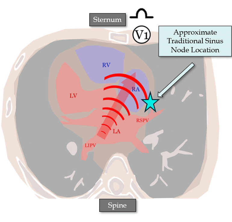
As we discussed last Beat Box, this can change, depending on whether the so-called “head” or “tail” of the sinus node is dominant, and where the depolarisation exits the epicardium and enters the endocardium.

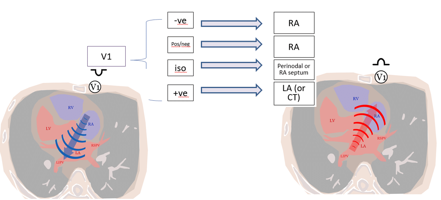
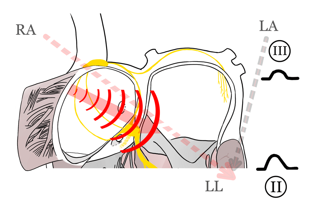
Lets look at how we can localise the Sinus Location, using the 12-lead P wave morphology.


Is the P wave origin RA or LA?

To determine this, we analyze V1:

A wavefront moving towards V1 produces a positive P wave, while one moving away from V1 produces a negative P wave.

  • Since the LA is the most posterior structure, Focal AT from the LA usually produces a positive P wave in V1.


  • A positive P wave in V1 can also indicate an SA nodal or Crista Terminalis origin in the RA, since both these anatomical regions are posterior.


Sinus P waves are classically biphasic (Pos/Neg) in V1 because:

  • Part of the wavefront moves anteriorly, depolarizing the RA towards V1.
    Another part moves posteriorly, depolarizing the LA away from V1.


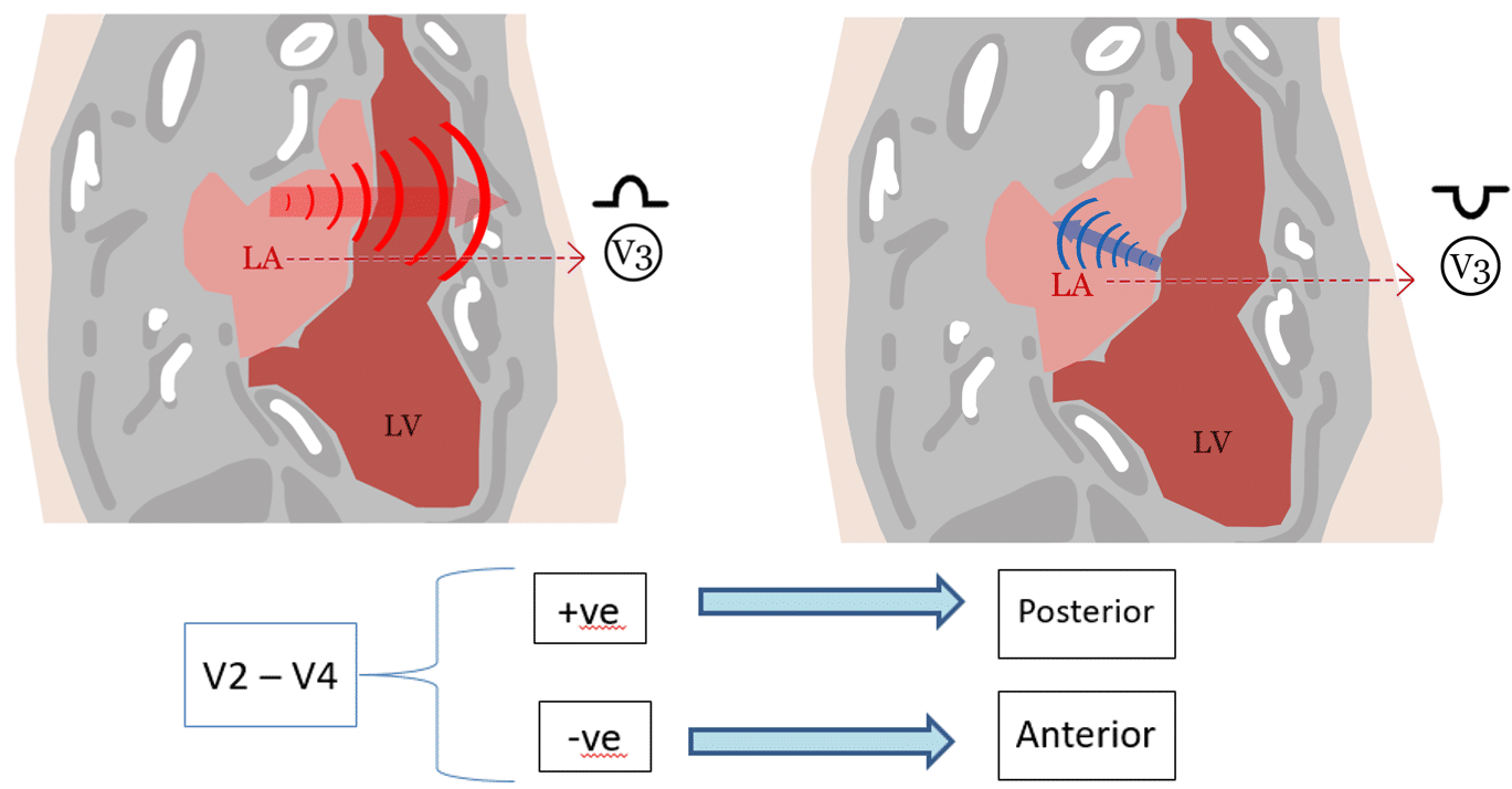
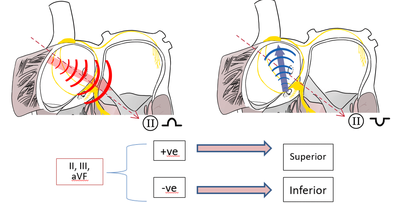
The diagrams below summarises how different atrial arrhythmia origins origins produce different P wave morphologies.


Is the origin Anterior or Posterior? – for this we analyse the P wave in V2-V4.

  • Posterior atrial sites, such as the SA Nodal or Crista Terminalis in the RA, or the pulmonary veins in the LA, tend to produce positive P waves in V2-V4.


  • This is because atrial depolarisation occurs towards the chest wall (where the precordial leads are located) producing a dominantly +ve P wave in these leads.


  • Since the SA node is located in a posterior site, it tends to produce positive P waves in V2-V4. 


Is the atrial focus coming from the top or the bottom of the atrium? – Look at the P wave in the inferior leads

  • Superior structures, such as the SA Node, SVC & the superior pulmonary veins result in a top –> bottom wavefront direction.


  • Since this moves WITH the inferior lead vector, this produces positive P waves in leads II/III/aVF.


Septal vs Lateral?

  • Lead II is more positive than lead III (higher positive amplitude) when the origin of tachycardia is located in a more rightward position.


  • This is generally seen in sinus ECG’s, since the SA Node is located in a very right lateral position.


  • Furthermore, Lead I is will produce a positive P wave when the origin is rightward located; since the lead 1 vector is positive when atrial depolarisation occurs in a right to left fashion.


My two cents:
So now, we’ve described what a sinus p wave looks like:

  • V1 is highly variable, it’s classically “pos/neg” but can also be just “pos” or just “neg”


  • V2-V4 = Pos, since the SA Node is a posterior structure


  • II/III/aVF = Pos, since the (Head of the) SA Node is a superior structure


  • May be “Neg” if the “Tail” of the SA node is active under vagal conditions.


  • I = Pos, since the SA node is a right lateral structure

Bonus tip = lead II is more positive than lead III, since the SA node is a right lateral structure.

We will revisit these P wave localisation principles when we dive into Focal AT ECG’s in future BeatBox posts. For those who want to read ahead, the below references are the seminal papers on P wave localisation.

Thanks for tuning in :)

Cheers

Mitch & CPiP Team.

References & Seminal papers on this subject:
Kistler PM, Chieng D, Tonchev IR, Sugumar H, Voskoboinik A, Schwartz LA, McLellan AJ, Prabhu S, Ling LH, Al-Kaisey A, Parameswaran R, Anderson RD, Lee G, Kalman JM. P-Wave Morphology in Focal Atrial Tachycardia: An Updated Algorithm to Predict Site of Origin. JACC Clin Electrophysiol. 2021 Dec;7(12):1547-1556. 

Kistler PM, Roberts-Thomson KC, Haqqani HM, Fynn SP, Singarayar S, Vohra JK, Morton JB, Sparks PB, Kalman JM. P-wave morphology in focal atrial tachycardia: development of an algorithm to predict the anatomic site of origin. J Am Coll Cardiol. 2006 Sep 5;48(5):1010-7. 

Leave a Reply

Subscribe to stay informed

Get the latest updates and event details, and be notified when new courses launch.