Busting Myths about Sinus Rhythm…

Question:

86yr female receives an ECG as part of a routine test. The question you receive: Is this sinus rhythm? Why are there inverted P waves?

(Click to zoom on image)

Answer:

Answer: The sinus node is bigger and more complex than often appreciated.

  • This is likely a “low” sinus rhythm/bradycardia.
    • Since the sinus node is exiting with an inferior origin, explaining the negative P waves in III/aVF and very positive P waves in aVL. The “low” sinus rhythm still has a posterolateral origin within the RA, which is why lead II is positive and V1 is the traditional “pos/neg” morphology associated with sinus rhythm and V2-V4 are all positive P waves.


  •  There is also a right axis deviation and possible LPFB and incomplete RBBB. But we are going to dive into the depths of “low sinus rhythm”.


Busting Myths about the Sinus Node being a “dot in the Right Atrium”

  • The SA node is a large cluster of pacemaker cells located on the lateral epicardial surface of the RA.


  • The “head” of the sinus node, is the densest cluster of automatic pacemaker cells at the SVC-RA junction.


  • The sinus node extends down almost the entire lateral wall of the RA, towards the IVC-RA junction where the “tail” of the sinus node is.


  • The tail of the sinus node contains another dense cluster of automatic pacemaker cells, however, these cells tend to spontaneously depolarise more slowly than the head of the SA node, partly due to gene expression and partly due to differences in autonomic inputs.


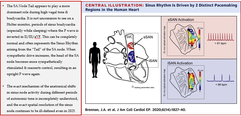
Brennan JA, Chen Q, Gams A, et al. JACC Clin Electrophysiol. 2020;6(14):1827–1840.


The size & architectural isolation of the sinus node from the rest of the atrial myocardium

Restrepo A, Razminia M, Sánchez-Quintana D, et al. J Am Coll Cardiol Case Rep. 2024;29

How do sinus impulses reach the atrium?

  • Since the SA node is electrically insulated from the atrial myocardium, it is necessary for sinus impulses to reach the atria via electrical bridges between the sinus node and atrial myocardium known as SA nodal extensions


  • These extensions are “exclusive” electrical connections which allow impulses to reach the atria, and they are found in somewhat consistent locations (although they are still poorly defined and possibly subject to variation).


These locations appear to the:

  1. The “traditional” SVC-RA junction point where you normally think of the sinus node being.
  2. The SVC
  3. The inferior lateral RA near the IVC
  4. A lateral-posterior exit beyond the crista terminalis
  5. The interatrial septum.


Clinical Relevance?
(Interestingly, when these “extensions” become diseased and non functional, we get SA nodal Exit Blocks, where the sinus node is disconnected from the atrial myocardium.)


Below, is an interesting video showing how sinus rhythm propagates through the endocardial aspect of the Right Atrium.

  • Notice, that there are several points in the atrium where the sinus impulse “suddenly appears from seemingly nowhere”.
  • These points appear to correlate with the known locations of the SA nodal extensions and likely represent endocardial breakthrough locations from impulses generated by the sinus node in the epicardium (which we cant detect on these maps).


My two cents:

  • Sinus rhythm can have inferiorly negative P waves, but the other leads should generally display traditional sinus P wave morphologies (lets dive into the what, how and whys of that on another day).
  • The Sinus node anatomy & it’s surrounding tissue is still ill-defined.
  • The so-called “tail” of the sinus node and the inferior sinus nodal exits appear to play a more dominant role in times of bradycardia due to differences in head & tail genetic expression and autonomic inputs.
  •  Even “Sinus Rhythm” can be so interesting!


Thanks for tuning in :)
Cheers
Mitch & CPP Team

References:

  1. Brennan JA, Chen Q, Gams A, Dyavanapalli J, Mendelowitz D, Peng W, Efimov IR. Evidence of Superior and Inferior Sinoatrial Nodes in the Mammalian Heart. JACC Clin Electrophysiol. 2020 Dec;6(14):1827-1840.
  2. Restrepo, A, Razminia, M, Sánchez-Quintana, D. et al. Myoarchitecture of the Sinoatrial Node and its Relevance for Catheter Ablation: Anatomy and Histology. J Am Coll Cardiol Case Rep. 2024 Jan, 29 (2).

Leave a Reply

Subscribe to stay informed

Get the latest updates and event details, and be notified when new courses launch.