Methods of unmasking Intramural Ventricular Substrate

Question:

VT Mapping has many complex techniques for identifying targets for ablation: S2 mapping, S3 Mapping, DEEP Mapping, Voltage Mapping, Isochronal mapping, LAVA, Late potentials, Conduction velocity.

Despite this, outcomes for VT ablation remain poor. Partially, this is due to poor characterization & ability to target intramural substrate. This Beat Box looks at methods of characterizing intramural substrate.

(Click to zoom on image)

Answer:

VT Substrate – intramural substrate is always relevant?

Ischaemic:

  • Ischaemic MI results in a predominance of subendocardial disease, however, there is often a significant amount of transmural disease penetrance with a 3-dimensional substrate of scar, adipose & surviving myocytes.


Non Ischaemic Cardiomyopathies:

  • Often has a greater predominance of intramural & epicardial scar, particularly in conditions such as Hypertrophic Cardiomyopathy, ARVC & Dilated Cardiomyopathy.


Bipolar Vs Unipolar mapping

  • Unipolar voltages are sometimes used as a surrogate for identifying epicardial/intramural scar because Unipolar EGMS are influenced by the depolarisation amplitude of the full thickness of myocardium.


  • Unipolar voltages are less dependent on wavefront direction with a wider field of view which may incorporate the full thickness of myocardial depolarisation.


  • Scar anywhere within the thickness of myocardium should conceptually lower unipolar voltage.


What cut offs to use for LV unipolar voltage?

  • Traditionally 3.3mV – 8.3mV.


  • Based on data collected in 5 structurally normal patients where >95% of EGMs demonstrated unipolar amplitude >8.3mV, with a 4mm point-by point mapping (low density) without contact force.


Intracardiac Echo

  • Regions of high echogenicity on Intracardiac Echo can also identify regions of scar.


  • ICE has the advantage of identifying intramural & epicardial scar as well as endocardial scar.


  • On ICE, myocardium that is thinned, akinetic, or has abnormal echo density can be delineated as scar, and has good correlation to scar defined by Electroanatomical mapping. These regions can be targeted for ablation.


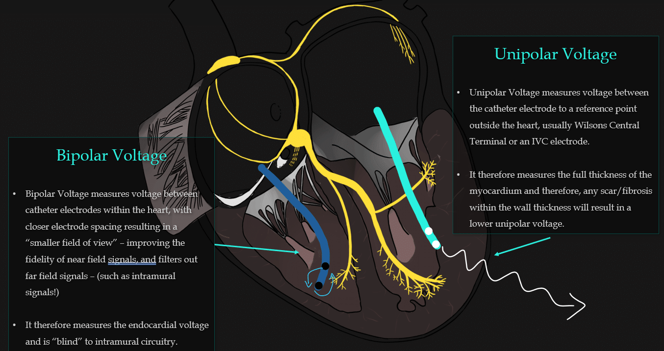
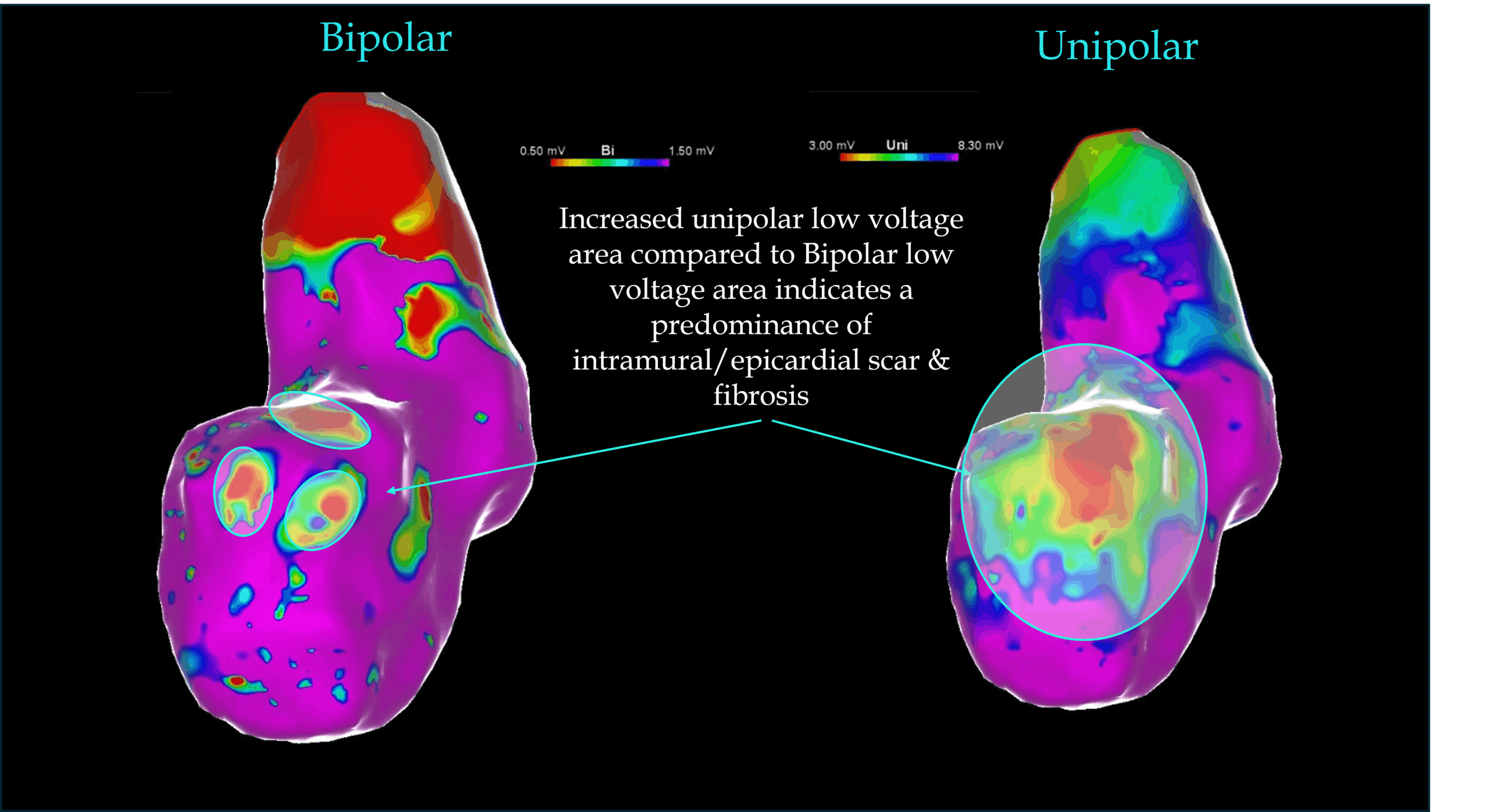
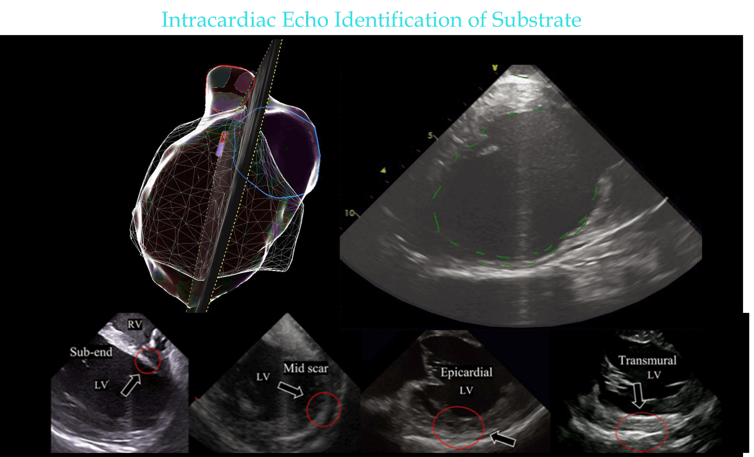
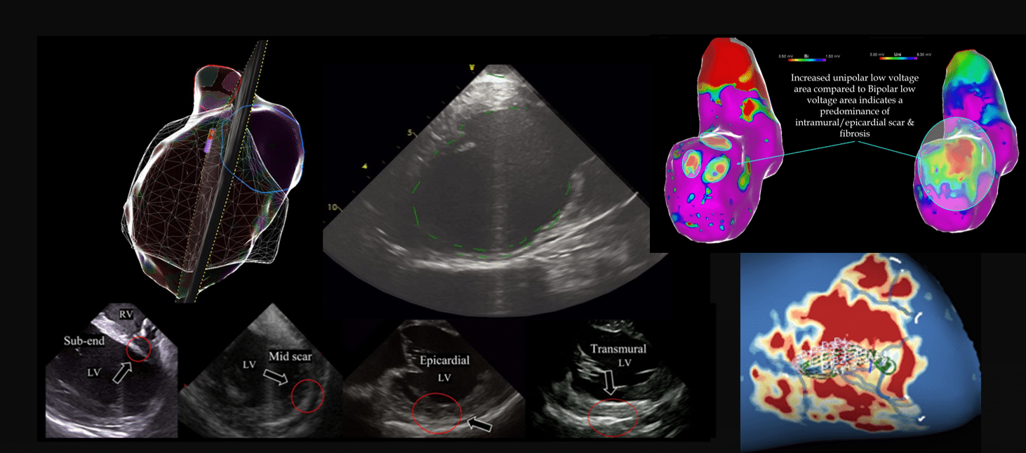
images modified from: Kanawati J, De Silva K, Bhaskaran A, Turnbull S, Zhou J, Kotake Y, Kumar S, Campbell T. Intracardiac echocardiography techniques to identify ventricular arrhythmia substrate. Heart Rhythm O2. 2022 Jun 17;3(5):602-612.

MRI identification of intramural fibrosis

  • MRI detects fibrosis using a Gadolinium contrast agent which distributes into the extracellular space, which is OVER-represented in fibrotic tissue, producing “hyper-intense” bright areas on MRI compared to normal myocardium.


  • Software such as the ADAS software can be used to identify fibrosis & regions of surviving myocytes located intramurally on MRI.


  • This enables the team to identify intramural substrate which may not be readily apparent using conventional mapping modalities.


My Two Cents:

  • Intramural substrate is always relevant, particularly in the context of non ischaemic VT etiologies, however, even ischaemic VT can heavily involve the mid-myocardial layers.


  • Conventional Bipolar mapping, EGM detection or ILAM mapping fails to detect critical intramural substrate.


  • Unipolar mapping can “suggest” that intramural disease may be present.


  • ICE can identify fibrosis at all layers of the myocardium and also monitor lesion creation in real time and give an indication of lesion intramural depth penetrance.


  • If available, MRI integration during VT ablation can offer additional resolution of intramural fibrosis, as detected by late gadolinium enhancement which is preferentially uptaken by fibrotic areas.


Thanks for tuning in :)
Cheers
Mitch & CPiP Team


This BeatBox post was based on the JET lectures contained in the Complex SVT Program 4, part of EP in Practice. EP Mastery in 2 years. No shortcuts. No compromises.

https://cardiacphysinpractice.com/ep-in-practice/


References:

  • Kanawati J, De Silva K, Bhaskaran A, Turnbull S, Zhou J, Kotake Y, Kumar S, Campbell T. Intracardiac echocardiography techniques to identify ventricular arrhythmia substrate. Heart Rhythm O2. 2022 Jun 17;3(5):602-612.


  • Nunes-Ferreira A, Brito J, Cortez-Dias N, da Lima da Silva G, Pinto FJ, de Sousa J. Preprocedural imaging guiding ventricular tachycardia ablation in structural heart disease. J Arrhythm. 2024 Dec 19;41(1):e13205.


  • Hutchinson M, Gerstenfeld E, Desjardins B, et al. Endocardial unipolarvoltage mapping to detect epicardial ventricular tachycardia substratein patients with nonischemic left ventricula cardiomyopathy.CircArrhytm Electrophysiol. 2011;4:49-55.


  • Hawson J, Al-Kaisey A, Anderson RD, Watts T, Morton J, Kumar S, Kistler P, Kalman J, Lee G. Substrate-based approaches in ventricular tachycardia ablation. Indian Pacing Electrophysiol J. 2022 Nov-Dec;22(6):273-285. doi: 10.1016/j.ipej.2022.08.002. Epub 2022 Aug 23.

Leave a Reply

Subscribe to stay informed

Get the latest updates and event details, and be notified when new courses launch.