
DEEP mapping – not without flaws

Question:

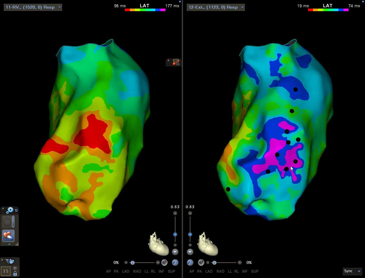
DEEP Mapping is somewhat fashionable in research and is making it's way into clinical practice. With images like below, it's hard to deny it's utility in unmasking substrate in VT ablation! So what is it, and what are it's shortfalls which are rarely talked about?

(Click to zoom on image)

Answer:

It’s utility – unmasking & quantifying functional substrate.

  • S2 mapping also unmasks functional substrate. But only DEEP mapping quantifies it. To understand “DecrEmental Evoked Potential” (DEEP) mapping, we first must understand S2 mapping.


S2 helps unmask functional substrate that is invisible to regular voltage maps.

The majority of VT substrate may be “functional” meaning that there’s not necessarily visible scar or fibrosis in regions associated with VT isthmus sites.

During VT, myocyte depolarisation occurs far more rapidly. Subtly diseased myocytes may exhibit a prolonged refractory, incomplete recovery between depolarisations and thus exhibit markedly slowed conduction velocity properties.

Theoretically, these “functionally diseased” regions may not exhibit unusual slowing or low voltage during sinus rhythm or S1 mapping, but may exhibit manifest disease during S2 or S3 mapping when under rate-related “stress”.

S2 mapping is used to uncover these regions of functional disease. DEEP mapping, is essentially quantifying the difference in Local Activation Time (LAT) between S1 and S2, theoretically allowing us to further demarcate the regions of “functional disease” that are not displayed on S1.


DEEP Mapping – unmasking VT isthmus sites?


  • DEEP mapping aims to uncover regions prone to “rate-related decrementation”.


  • Regions prone to rate related decrementation have been shown to be a stronger correlate to VT isthmus sites than late potentials alone.


  • DEEP mapping may be useful in showcasing potential ablation sites.


Below, you can see that DEEP mapping appears to “unmask” potential VT substrate that remained hidden with traditional mapping modalities. 


Jackson et al found DEEP to correlate more strongly with VT isthmus sites than late potentials.


DEEP Mapping shortfalls

1. Directionless mapping

  • Not really a problem, but DEEP mapping provides no information on wavefront direction. It only says “the EGM at this location occurred later on S2 vs S1”.
    • The S2 isochronal map can be used to interpret wavefront direction alongside the DEEP map, so this is not really as problem, but still worth pointing out.


2. Potential for a “shadowing effect” & overestimating disease.

  • DEEP mapping says “the EGM at this spot occurred later on S2 vs S1”. That statement is not the same as saying “the tissue at this spot decremented on S2”. Because maybe the EGM at this location occurred later because the proximal tissue (between “this” EGM and the pacing site) decremented.


  • DEEP maps arrival time, which is a cumulative sum of conduction delays along the path from the pacing site — not a local conduction property. If “decrement” occurs 2cm up stream, then every single EGM downstream will show a later EGM, even if the downstream sites have completely healthy tissue.


  • This can sometimes (though not always in my experience) produce a “shadowing effect”, where healthy regions located behind an area of disease are more likely to show up as “decremental”, even though they are healthy. This can lead to an overestimation of substrate.


3. We still miss intramural/epicardial substrate.

  • DEEP mapping is a bit of a mis-nomer. It doesn’t map “deep” at all. It’s still a primarily surface mapping method.


4. Inconsistent EGM annotation

  • Annotating EGM’s during DEEP mapping can have huge inter and intra-operator variability. For example, the EGM below, what would you annotate? The offset of the Bipolar EGM? The largest deflection?


  • You may be able to improve consistency by steadfastly annotating on the steepest dv/dt of the unipolar. But to be honest, I’m still uncertain if that’s the right thing to do in practice. Often there are little unipolar “blips” at the end of the S2 bipolar EGM, that you are tempted to annotate to, despite it not being the “steepest” part of the unipolar signal. I think there’s not a whole lot of consensus on this, and there’s much interlab variation.


  • Annotation inconsistency is somewhat corrected by automated annotation developed by our friendly 3D mapping companies. However, from experience sometimes the nicest juciest signals are missed by these algorithms. They might filter out those unipolar “blips” or annotate a fractionated EGM in the middle, completely missing its value in a sea of 10,000 data points.


  • There is still a very very important role for the mapper, tech & physician manually visualising EGMs.


My Two Cents:

  • DEEP mapping quantifies the decrement between S1 and S2, helping unmask possible functional substrate.


  • DEEP mapping unfortunately does not map “local disease” but rather the arrival time of the EGM in a location. That arrival time can be delayed by either disease at the location, or by disease between the location and the pacing site causing delay.


  • Because of this property, local disease can sometimes create a “shadowing effect” by delaying the EGM arrival of distant tissue, lowering the spatial resolution of the technique.


  • DEEP mapping still misses all the “deep” substrate -it only maps the surface.


  • It’s another tool in the toolbox, and best interpreted alongside S2 isochronal mapping, & ICE for a more complete picture of VT substrate.


Thanks for tuning in :)
Cheers
Mitch & CPiP Team


This post was based on a Mini-Lecture from the Scar VT Program of EP in Practice. No Compromises. No Shortcuts. Built for those who believe in more than the minimum standard.

EP in Practice – Product Overview


Reference:

Jackson N, Gizurarson S, Viswanathan K, King B, Massé S, Kusha M, Porta-Sanchez A, Jacob JR, Khan F, Das M, Ha AC, Pashaei A, Vigmond E, Downar E, Nanthakumar K. Decrement Evoked Potential Mapping: Basis of a Mechanistic Strategy for Ventricular Tachycardia Ablation. Circ Arrhythm Electrophysiol. 2015 Dec;8(6):1433-42.

Leave a Reply

Subscribe to stay informed

Get the latest updates and event details, and be notified when new courses launch.