The little appreciated RV infarction: Detection & Consequences

Question:

74-year-old male with a history of ischemic heart disease presents with resting central chest heaviness & hypotension. What does the ECG show? Is there cause for concern?

(Click to zoom on image)

Answer:

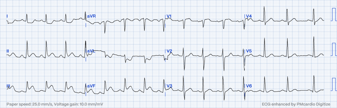
Sinus rhythm with inferior MI & likely RV involvement. This patient had an occluded proximal RCA which was treated with emergency PCI.


Detection & explanation

ST elevation in leads II, III, aVF & V1, with high lateral depression (I & aVL)

  • V1 sits on the right side of the chest, closest to the RV region. ST elevation here & in the inferior leads, with reciprocal depression in the high lateral leads (I & aVL), is characteristic of an inferior LV wall MI with concurrent RV infarction. This usually indicates a proximal Right Coronary Artery (RCA) occlusion, as a dominant RCA supplies the inferior wall of the LV as well as the RV via the RV branch (usually originates from the mid-RCA). Interestingly, the ST elevation in V1 can be subtle, as it may be cancelled out by reciprocal depression from inferior ST elevation.


  • Another characteristic of RV MI is lead III ST elevation > lead II in combination with the above ECG features.


The Right Ventricle is supplied by the RV marginal branch which branches off the mid RCA in most cases. This means that proximal or mid-RCA occlusions can also result in RV infarction & occur in approximately one-third of patients who have an inferior MI (of the LV).


Pathophysiology of RV infarction:

  • Infarction of the Right Ventricle is associated with an almost 3-fold increase in mortality, compared to inferior MI without RV infarction.
  • Partially, this is because an RV MI usually indicates a proximal RCA occlusion, meaning that an extensive territory of the LV myocardium is also injured.
  • Furthermore, resulting RV dysfunction can contribute to hypotension as it fails to propel blood forward into the pulmonary system, depriving the left ventricle of filling pressure & volume.
  • The resulting increase in RV and RA diastolic pressures causes acute RV dilatation, displacing the interventricular septum leftwards , further limiting LV filling (which is already volume deprived), and exacerbating systemic hypotension, and further diminishing coronary perfusion.
  • This can be lethal if not managed appropriately & is associated with more ventricular arrhythmias, AV block & haemodynamic collapse compared to inferior MI without RV involvement. (Graph below).

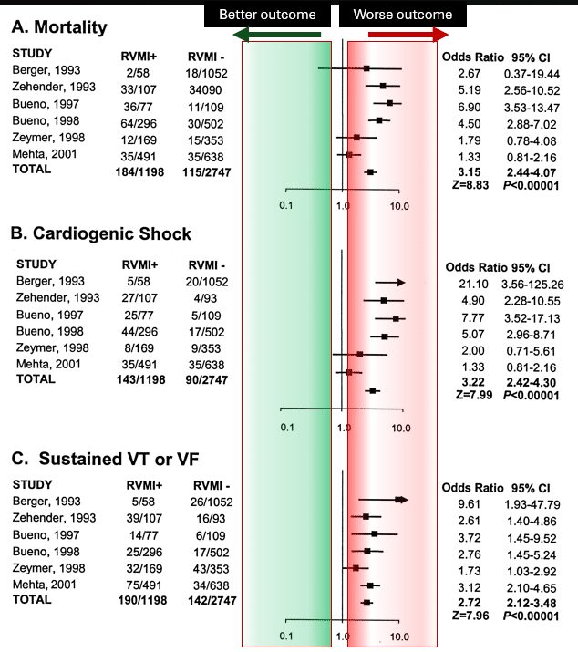
Prognosis of inferior MI with RV involvement vs no RV involvement 

My two cents:

Inferior MI with concurrent RV infarction is a serious diagnosis with an almost 3-fold increase in mortality & is associated with a large territory of injury. The physiology of RV MI, predisposes to haemodynamic collapse & resulting brady & tachyarrhythmias. Concurrent RV infarction can most often be suspected when inferior ST elevation occurs concurrently with V1 elevation & high lateral depression, with lead III ST elevation > lead II.

Thanks for tuning in :)
Cheers
Mitch & CPP Team


More on inferior LV & RV MI can be found in ECG in Practice Program 1
https://cardiacphysinpractice.com/ecg-in-practice/

  • Hamon M, Agostini D, Le Page O, Riddell JW, Hamon M. Prognostic impact of right ventricular involvement in patients with acute myocardial infarction: meta-analysis. Crit Care Med. 2008 Jul;36(7):2023-33.
  • Somers MP, Brady WJ, Bateman DC, Mattu A, Perron AD. Additional electrocardiographic leads in the ED chest pain patient: right ventricular and posterior leads. Am J Emerg Med. 2003 Nov;21(7):563-73.
  •  Mehta SR, Eikelboom JW, Natarajan MK, Diaz R, Yi C, Gibbons RJ, Yusuf S. Impact of right ventricular involvement on mortality and morbidity in patients with inferior myocardial infarction. J Am Coll Cardiol. 2001 Jan;37(1):37-43.

Leave a Reply

Subscribe to stay informed

Get the latest updates and event details, and be notified when new courses launch.