Hidden Dangers: When Innocent ECG Features Turn Hazardous Together

Question:

81-year-old female with history of sudden fall with facial injury. Describes a history of multiple dizzy spells when walking. After her fall, she presents by ambulance to hospital. This is her ECG. Is there concern of a cardiac cause?

(Click to zoom on image)

Answer:

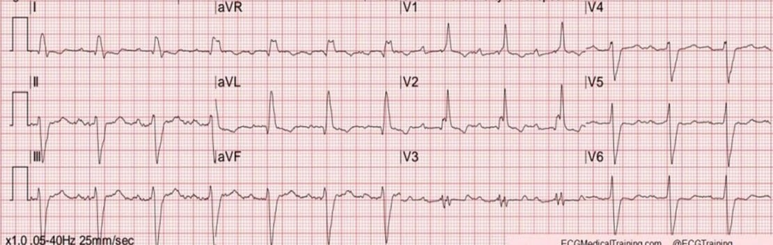
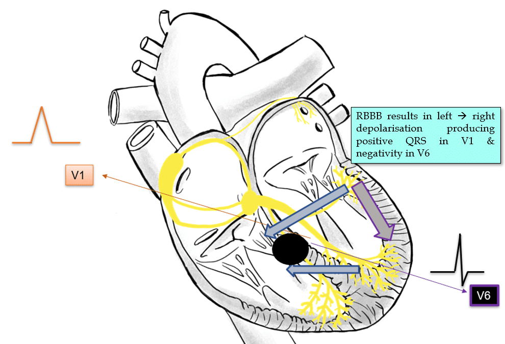
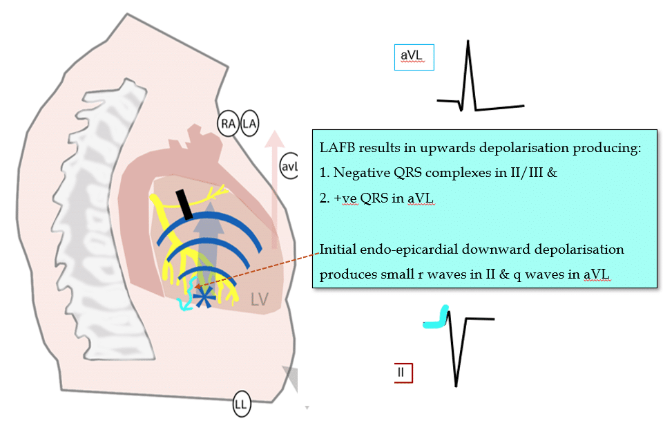
Answer: Sinus rhythm with RBBB, Left axis, 1st degree AV conduction delay & Left Anterior Fascicular Block. (So-called “Trifascicular block” – please don’t use that term. It’s misleading).

Evidence of disease in multiple locations, impacting the function of the cardiac conduction system. Given her history of falls & dizzy spells, there are strong reasons to be concerned about the possibility of intermittent conduction system failure (resulting in complete heart block, or ventricular standstill or high grade 2nd degree AV block) causing her symptoms.


Her ECG features of note are:

  • 1st degree AV conduction delay
    • (PR >200ms)
    • Indicative of slow conduction between the atria & ventricles. In isolation, this is rarely a cause for concern and is usually the result of high vagal tone and is completely reversible. However, in the context of multiple conduction system defects on ECG & the patient’s symptoms, there is some concern that this delay could be the result of a diseased His-Purkinjie system.


  • RBBB
    • (rSR complex V1-V3 with QRS width >120ms in V1, slurred prominent S waves V5/V6 with duration >50% of QRS complex reflecting delayed RV depoalrisation).
    • Again, in isolation, Right Bundle Branch Block is rarely a cause for immediate concern. However, in the context of syncope & the other conduction system defects (like LAFB), it hints at the possibility of an irriversibly diseased His-Purkinjie system.

  • LAFB
    • (Left axis deviation, rS pattern in II/III – the “r” comes from initial endocardial to epicardial depolarisation, qR pattern in aVL & R wave peak time >45ms aVL).
    • Once more, in isolation LAFB is very rarely cause to bat an eye lid. However, in the context of additional conduction system defects (such as RBBB) and a history of syncope, LAFB can make the difference between implanting a pacemaker, or sending the patient home.

My two cents:

Bi-fascicular block with 1st-degree AV conduction delay (often called “trifascicular block”) in the context of unexplained syncope strongly suggests a cardiac cause due to the high likelihood of severe His-Purkinje disease.

  • A thorough history of symptoms is needed. Syncope with facial fracture is particularly concerning, as vasovagal episodes rarely result in facial injury.
  • The patient’s ECG features indicate right bundle branch, left anterior fascicle, and possibly AV nodal disease. The left posterior fascicle appears unaffected, but this absence of evidence doesn’t rule out disease & intermittent conduction system failure resulting in haemodynamic collapse is a probable cause for this patients symptoms.


These findings warrant further investigation with an EP study to assess the His-Purkinje disease burden or consider pacemaker implantation.

Cheers
Mitch & CPP Team

P.s. This ECG was actually from a 77 year old male. I realised after I posted the blog. It doesn’t change the lessons learnt, but I wanted to set the record straight for the sake of correctness :)

Leave a Reply

Subscribe to stay informed

Get the latest updates and event details, and be notified when new courses launch.