Dropped P waves in the dark. Cause for concern?

Question:

The following was a holter monitor strip taken from a 74 year old man with a history of unexplained syncope & occurred at 01:20am whilst asleep.

What is the rhythm & is this cause for concern?

(Click to zoom on image)

Answer:

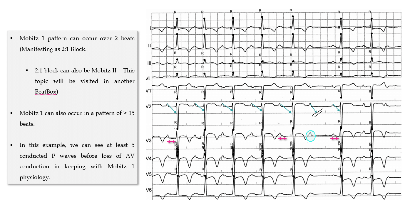
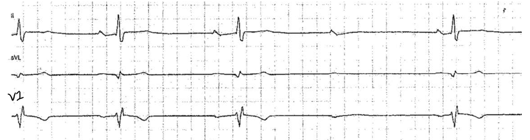
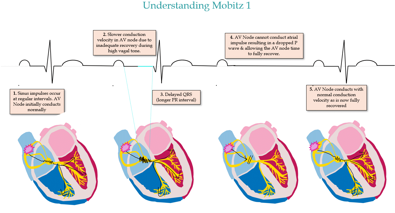
Answer: Sinus Rhythm with 2nd degree AV Block, Mobitz 1, driven by high parasympathetic (vagal) tone.


ECG Features of Mobitz 1 AV block

  1. Progressively lengthening PR interval followed by a non conducted P wave.

  2. The subsequent conducted P wave will then have a PR interval that is shorter than the last conducted PR interval prior to the non conducted beat.


Why It Happens:

  • Mobitz 1 typically occurs due to high parasympathetic drive slowing function of the AV node (such as during sleep – this is normal and may happen to you at night!). It normalises with increased sympathetic activity, like exercise.


  • This phenomenon is a result of the AV node’s inherent decremental conduction properties and sensitivity to autonomic tone.
    • The AV node is densely innervated by sympathetic and parasympathetic inputs.
    • The conduction velocity and repolarisation time of the AV node varies significantly in proportion to fluctuations in autonomic tone.
    • High vagal tone/parasympathetic drive will slow conduction velocity through the node.


Physiology Breakdown:

  • Parasympathetic drive slows the AV node’s conduction velocity, & repolarisation time increases.


  • With every sinus beat, the AV node’s recovery process begins to “fatigue,” leading to progressively slower conduction with every sinus beat, resulting in progressively longer PR intervals.
    • In Mobitz 1, AV nodal decrementation is characterized by reduced calcium channel recovery between consecutive atrial impulses. This results in a slower phase 0 depolarization and progressively slower conduction through the AV node.


  • Eventually, the AV node can’t conduct the impulse due to it being fully refractory—resulting in a non-conducted P wave. Importantly, this allows the AV node time to recover, meaning that it is fully capable of conducting the next sinus beat (with a relatively short PR interval).


Clinical Significance:

CONTEXT is everything:

  • Mobitz 1 is usually benign during periods of high vagal tone & represents normal human physiology. It is entirely reversible, usually haemodynamically stable & occurs within the AV node itself due to high vagal tone and is rarely correlated with His-Purkinjie conduction system disease.
    • I most commonly see this in resting athletes, sleeping patients (sometimes exacerbated by antiarrhythmics), patients on a Tilt Test experiencing a vasovagal episode, & patients who have an acute inferior MI resulting in high vagal tone (see this post for more detailed pathophysiology on this!)


  • It is strange, worrying & maladaptive if observed during high sympathetic tone, such as during a treadmill test!


  • Mobitz 1 typically resolves with an increase in sympathetic tone (think: patient agitation or movement). In rare cases where symptoms persist, a pacemaker might be considered, but that’s more of an exception than the rule.


My Two Cents:

  • Mobitz 1 is the benign brother of it’s more sinister sibling Mobitz II AV block, classically presenting as progressively increasing PR intervals, followed by a non conducted P wave.
  • It represents “normal” human physiology and is most often caused by high vagal tone during sleep or relaxation.


    • In EP studies, we actually “force” this sort of AV block to occur in every single normal patient, as a matter of routine. It’s a very normal response of the AV node in the context of high vagal tone. 


    • It would be very “abnormal” to see Mobitz 1 occur in the context of high sympathetic tone, such as on a treadmill. Mobitz 1 would be a strange, worrying and incredibly maladaptive response to exercise. 


  • You commonly see this in Holter monitors during sleep (its quite possible you have Mobitz 1 at 01:20AM as well!) or in resting Athletes ECG’s who usually have high baseline vagal tone.
  • It is rarely cause for concern unless causing symptoms (whereas Mobitz II is rarely NOT cause for concern – more on that in a future Beat Box).


Thanks for tuning in :)
Cheers
Mitch & CPP Team

Learn more about Bradyarrhythmias in our ECG in Practice – Program 1


Another example of Mobitz 1:

Leave a Reply

Subscribe to stay informed

Get the latest updates and event details, and be notified when new courses launch.